
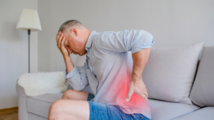
Failed back surgery syndrome (FBSS) is the term for a patient’s continued pain after an unsuccessful back surgery. The patient is usually referred to a pain management specialist to treat pain.
The goal of spinal surgery is to decompress a nerve root or to stabilize a painful joint. FBSS occurs when surgery is unsuccessful or negatively affects a structure near the problem area.
Spinal surgery is complicated because it can be hard to diagnose the cause of the pain. X-Rays and MRIs limit what doctors can see because the pain often occurs when the patient is moving and active. Imaging only provides still images.
Failed Back Surgery Syndrome: Signs, Symptoms, and Treatment
Signs & Symptoms of FBSS
One common complaint physicians hear from patients is chronic back pain. However, not everyone experiences the same pain, and the type of pain they experience can vary, based on their spinal disorder, their previous procedure, or underlying cause of FBSS.
Other types of pain that can be associated with FBSS are:
- Nociceptive Pain – Localized pain that may be dull or sharp.
- Neuropathic Pain – Nerve-related pain is caused by nerve or spinal cord damage.
- Radicular Pain – Radicular pain radiates from one area to another
Other common symptoms of Failed Back Surgery Syndrome include:
- Return of original symptoms & pain – When the symptoms return that the original procedure was intended to correct, it may be a sign of FBSS.
- Reduced mobility – Recovering from back surgery is a process. However, if mobility is reduced or limitations arise that are different than predicted by your physician, it could be a sign of FBSS.
- New problems arise – While the surgery may correct the original symptoms, new pain in a different part of the spine should be discussed with your doctor.
- Onset of Headaches – If headaches were not a part of your medical history prior to having surgery, this may be a sign of nerve damage after a spinal procedure.
Diagnosing Failed Back Surgery Syndrome
The first step to diagnosing FBSS pain is to see a doctor for a physical exam. Motion, sensitivity, and strength help doctors decide the next steps to correctly diagnose the pain. Your medical history, pain levels, and previous treatments are discussed with your doctor to help them define the pain and decide on a treatment method.
Having tests like CT scans, MRIs, and X-Rays help doctors take an inside look at your pain. The images don’t always show the root cause of the pain, making FBSS hard to diagnose.
Since imaging is not always reliable, doctors may use another form of testing to diagnose your pain. This is called an aware state surgical examination. A doctor uses a small probe and stimulates the spinal cord, mimicking the pain, and triggering a response from the patient. This informs the doctor what type of pain the patient has and how it is caused.
Back pain patients are referred to pain management specialists who treat their pain and help improve their quality of life through short-term techniques.
Conservative Treatments for Failed Back Surgery Syndrome
There are many conservative treatments for FBSS.Besides the physical therapy and pharmacological therapy with over the counter or prescription medications, doctors use minimally invasive interventional modalities such as epidural steroid injections and injections of the lumbar facet joints or associated medial branches, with or without radiofrequency ablation rhizotomy (RFA).What makes these a consrvative first choice option is the minimally invasive nature of the procedures.These therapeutic interventions are designed to treat the pain and provide temporary symptomatic relief.
Neuromodulation in the Treatment of FBSS
In the treatment of Failed Back Surgery Syndrome, one of the most successful treatment options is Spinal Cord Stimulation. Treatment efficacy for FBSS has increased over the years with the majority of patients experiencing pain relief and reduced medicinal load. Improved quality of life can also be achieved using SCS.
For patients with unrelenting back pain due to mechanical instability of the spine, degenerative disc disease, spinal injury, or deformity, spinal surgery is a well-accepted treatment option; however, even after surgical intervention, many patients continue to experience chronic back pain that can be notoriously difficult to treat. Clinical evidence suggests that for patients with FBSS, repeated surgery will not likely offer relief. Additionally, evidence suggests long-term use of opioid pain medications is not effective in this population, likely presents additional complications, and requires strict management.
SCS has been shown to be a safe and efficacious treatment for this patient population. Recent technological developments in SCS offer even greater pain relief to patients’ refractory to other treatment options, allowing patients to regain functionality and improve their quality of life with significant reductions in pain.
To learn more about FBSS and how the team at Progressive Pain Management can help treat your pain, fill out the form below.
Facet joints are small, bony joints that sit along the spine. They are paired up from the neck to the lower back area. When the joints get inflamed, they cause pain that can be severe. The pain can be acute or chronic, caused by a variety of conditions.
The pain radiates to other parts of the body and causes indirect pain. For pain that starts in the lower back, it’s common for the pain to move to the thighs and buttocks. If the pain starts in the neck or upper back region, it can transfer to the shoulders, and arms, and even cause headaches.
Facet Joint Injections for Chronic Pain
What is a Facet Joint Injection?
The injection is a strong anti-inflammatory steroid that gets inserted into the facet joint. The medication is a mixture of a local anesthetic and a steroid, similar to a cortisone injection. Before performing the injection, your doctor will explain the procedure, have you sign all the appropriate consent forms, and then have you lie on your stomach.
The procedure is minimally invasive. X-ray guidance is used to locate the facet joints and helps guide the needle to the exact location. The actual injection only takes a few minutes, but you may want to allow for additional recovery time depending on the type of anesthetic you choose. Some doctors will let you choose a local anesthetic or a stronger general anesthetic. The general anesthetic may cause drowsiness and sedation will take longer to wear off.
What to Expect After the Injection?
Right after the injection, you will feel relief from pain. That is due to the anesthetic in the injection, and only lasts for a few hours. The injection site may become sore after a few days, and the pain is not uncommon to resume the day after the injection. About 1-2 days after the injection, the medication will begin to take effect and help reduce your pain on a daily basis.
It is advised to take it easy following the injection and only to resume normal activity when it is comfortable to do so. Your doctor may recommend applying ice to the injection site. This will help reduce any swelling and soreness following the procedure.
How Long Does a Facet Joint Injection Last?
Immediately following the injection, you may feel lessened pain. However, this is temporary relief until the medication kicks in. That can take between 2-7 days after the injection.
If the first injection is successful in managing your pain, your doctor may perform additional injections. The amount of relief depends on your specific pain and because there are a variety of pain receptors in the spine, the long-term effect of these injections cannot be predicted. Everyone’s pain is different, so the treatment effectiveness will vary.
There are very few risks associated with this type of injection. The most common being tenderness and pain at the injection site, although this is temporary. Side effects of steroids may include weight gain, water retention, and temporary increase in blood sugar.
Facet joint injections are an excellent alternative to undergoing serious back surgery. Talk with your doctor today to discuss your options for getting relief from your pain and taking one step closer to bettering your quality of life.
Risks Associated with the Facet Joint Injection Procedure
As with any type of injection or invasive procedure, there are certain risks or complications that could arise:
- Allergies: In most cases, the allergic reaction is caused by the X-ray contrast or steroid, not the local anesthetic. Severe allergies are rare.
- Bleeding: In patients who are taking blood thinners or have a bleeding disorder, it is possible to have some bleeding as a result of the injection.
- Discomfort at the Injection Site: Worsened pain or discomfort at the injection site. Long-term pain is uncommon.
- Infection: Minor infections occur in less than 2% of facet joint injections. More complex infections are rare, only happening in 0.1% of injections.
Fill out the form below to contact the Progressive Pain Management team and learn more about facet joint injections.
Spinal cord stimulation therapy is a pain treatment that masks the pain signal before they reach the brain. A device similar to a pacemaker is implanted in the body and delivers electrical pulses to the spinal cord. This is an option for patients with chronic, leg, or arm pain.
What is Spinal Cord Stimulation?
A spinal cord stimulator (SCS) is a small device that is placed under the skin and transmits a mild, low-frequency electric current to the spinal cord. A tiny wire transfers the pulse to the nerve fibers. The SCS minimizes pain because the current modifies and hides the pain signals from reaching the brain.
Spinal Cord Stimulation for Chronic Pain
It is important to note that spinal cord stimulation therapy does not get rid of the source of the pain. It simply runs interference with the signal to the brain. This means that pain relief can vary depending on the patient. The SCS device produces a slight tingling sensation.. It is this sensation that overrides the pain signals. Pain signals travel on the small nerve fibers, whereas the fabricated signals from the SCS travel on larger, more dominant nerves fibers.
The goal of spinal cord stimulation is not to completely erase pain, but to provide a 50-70% reduction. Even the slightest bit of pain relief can be helpful to someone who suffers regularly. Before a permanent spinal cord stimulator is implanted, each patient undergoes a trial to make sure this type of therapy will be effective and reduce their pain.
Why is SCS Used?
Spinal cord stimulation is used to treat neuropathic pain. This is pain that originates from nerve damage. The nerve damage could be caused by injury, accident, or trauma. Patients who are prime candidates for SCS have typically suffered from chronic pain in the lower back, leg, or arm. Commonly, these patients have also had previous surgeries.
More frequently, SCS is being used to avoid back surgery. Other leading causes for receiving SCS therapy is complex regional pain syndrome and peripheral neuropathic pain. Nerve pain that spans beyond damage to the brain and spinal cord, such as from an infection or even amputation or diabetes, is another reason that SCS may be recommended by your physician.
More recently, SCS therapy has been proven to treat a number of chronic visceral pain types, such as abdominal or pelvic pain.
Spinal cord stimulation therapy is used when other treatment types have not been effective in reducing chronic pain or if the patient does not want to undergo surgery. Fortunately, there are no pre-existing medical conditions that would prevent someone from receiving this type of therapy. If you have pain that is caused by a correctable problem (meaning it could be fixed by having surgery or other interventional treatments), SCS is a viable option for reducing your pain.
This type of therapy is more effective when utilized in the earlier stages of a chronic disease or condition, rather than later when a disability has been established.
SCS therapy is used to reduce these types of pain:
- Failed Back Surgery Syndrome: When initial surgery (or surgeries) have been ineffective in reducing pain on a consistent basis.
- Sciatica or Arm Pain: Persistent pain caused by arthritis, spinal stenosis, or extensive nerve damage.
- Complex Regional Pain Syndrome: When patients experience severe chronic pain, typically in their hands or feet.
- Arachnoiditis: This is painful inflammation and scarring of the protective lining of spinal nerves
Other types of pain caused by stump pain, peripheral vascular disease, multiple sclerosis, or a spinal cord injury may be reduced by the use of a spinal cord stimulator.
Benefits of Spinal Cord Stimulation
Spinal cord stimulation therapy reduces the number of abnormal pain signals from reaching the brain. However, it also helps the body restore pain-inhibition pathways that have been lost. Pain-inhibitory pathways essentially work as a gate-keeper. They control how much pain is received by the brain. SCS therapy harnesses the body’s natural pain-relieving chemicals that are used by nerve fibers to communicate with each other. Not only does this whole process reduce pain, but it increases microcirculation.
It is reported that 50-70% of patients who are candidates for SCS therapy experience 50% reduction in pain. An even higher proportion can expect to experience a 30% reduction in pain levels. For many patients who suffer from chronic pain, even the smallest amount of pain relief is welcomed. This has a profound effect on improving the quality of life in patients who have suffered from long-term chronic pain.
Learn more about spinal cord stimulation and how it works for treating chronic pain, fill out the form below and get in touch with the team at Progressive Pain Management today.
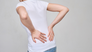
Sciatica causes pain to radiate along the sciatic nerve’s path. It can disturb daily activities, quality of life, and causes discomfort. Pain can be caused when the nerves get pinched, and intense pain can travel into your legs. Sciatica can be repetitive, intolerable, and many times, the cause is unknown to the patient.
This type of pain can be treated with self-care and rarely requires medicine. Treatments depend on what is causing the underlying root of the pain. Natural remedies are available and can dramatically improve a patient’s condition. Patients should always be discussed with a medical professional before beginning any type of treatment.
Natural remedies for sciatica
5 Natural Remedies for Sciatica Pain
Chiropractor
Patients who suffer from sciatic nerve pain often find relief from chiropractic adjustments. Adjustments can improve spinal function and in turn, reduce pain.
Yoga
While sometimes moving can aggravate sciatic pain, certain types of movements and stretching can actually reduce pain. Stretches that lengthen the spine or yoga positions that help develop good posture help reduce stiffness, inflammation, and pain.
Yoga is safe and effective for people with sciatic pain. It can strengthen core muscles, relax stiff areas, and help prevent further pain.
Acupuncture
Acupuncture is a traditional Chinese practice that maintains health by releasing the body’s natural energy. Tiny needles are injected into specific, targeted areas. Acupuncture helps improve blood flow, oxygen, and channels energy in a holistic approach. It has been FDA approved to treat back pain and chronic pain of all kinds, including sciatica.
Active Lifestyle
Working at a desk or lounging too long can worsen back pain. Sciatica treatments frequently recommend movement and staying active. An active lifestyle and targeted exercises allow for joints and affected areas to loosen up and relieve pain. Isometric exercises help relieve pain during the day; many of these can be performed at work or on the go. If you typically lead a somewhat sedentary lifestyle, investing in a fitness tracker can help motivate you to set activity goals.
Heating Pads
Heat loosens tight muscles and promotes blood circulation. Many people with sciatica find pain relief by using heating pads on a low setting for 15-20 minutes daily. Applying heat to the affected or painful area can be performed a few times a day, every few hours. Many heating pads are small and travel well, so it can be helpful to use while sitting at work. Another method of heat is taking warm baths. Water relieves pressure from your joints, also reducing pain, while the heat loosens up the muscles. For use on-the-go, one-time-use heating wraps may be used that are effective for several hours.
If you suffer from sciatica pain, the team at Progressive Pain Management can help. Fill out the form below to get in touch.
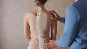
Degenerative adult scoliosis, also known as adult-onset scoliosis, is when the facet joints and intervertebral discs deteriorate and lead to a curve in the spine. This side-to-side curve occurs over time as a person gets older. It commonly begins after age 40, and is more frequently seen in patients who suffer from osteoporosis. Osteoporosis causes the bones to lose bone density, quickening the deterioration rate. The spine can begin to “lose shape,” and a scoliotic curve can develop.
What is Degenerative Scoliosis?
Causes of Degenerative Scoliosis
Age is the leading cause of degenerative scoliosis. Over time, facet joints and discs can deteriorate over time, causing the spine to weaken and lose shape. The joints and discs are what make your back flexible. The joints enable bending and twisting. The discs act as shock absorbers.
Degenerative changes are all part of getting older; however, there are other conditions that cause degeneration at high rates:
- Arthritis: Weak joints and irregular vertebral alignment caused by pressure on the spine.
- Osteoporosis: Loss of bone density weakens the bones and their stability.
- Spine Surgery: Recovery and time after a spinal surgery can cause spine deformities.
Aging joints, combined with any of the factors above, can cause significant deformity. Mild-to-moderate deformity is common in older people but can appear in people as young as their 40s. Studies show that approximately 60% of people over the age of 60 have mild degenerative scoliosis.
Treatments
Degenerative adult scoliosis can cause pressure on the nerves and along the spinal cord. This leads to weakness, numbness, and pain in your lower extremities. In extreme cases, the pain and weakness can make walking difficult.
Unlike scoliosis in adolescents, treatments for spine deformity in adults is determined by the severity of the symptoms, not the severity of the curve of the spine. Treatments tend to focus on managing the pain and reducing symptoms as opposed to correcting the curve.
For mild-to-moderate cases, treatment usually consists of pain management and physical therapy (PT). PT is used to increase mobility of the spine. Low-impact exercises may be used to help restore strength of the spine, with hopes of decreasing the symptoms and rate of deterioration. Over-the-counter medications can be used to manage and reduce pain. NSAIDs help minimize pain caused by inflamed joints or arthritis. Medications are not used to heal the spine, but to reduce pain caused by the curvature of the spine. Reducing pain can allow a patient to resume normal daily activities while they work to regain strength and flexibility.
Other ways to treat degenerative adult scoliosis include:
- Weight Loss: Losing weight helps decrease the pressure placed on facet joints
- Nutrition: Eating foods that help reduce anti-inflammatory properties – such as tumeric and ginger – help reduce the pain. Drinking water is important
- Bracing: A brace is used to restrict motion to relieve pain during daily activities and perhaps during physical therapy. Braces can also be used to decrease stress on the facet joints.
If the symptoms and curvature of the spine is severe, your doctor may recommend surgical options. Surgery is used when the patient’s pain is so severe that is become debilitating or when there is nerve damage from the spine curving. Nerve damage can result in the loss of normal bodily functions such as walking or bladder control.
To learn more about degenerative adult scoliosis, contact the team of experts at Progressive Pain Management. Fill out the form below to get in touch today.
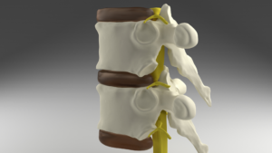
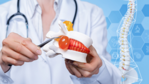
Discs are the cushions between the vertebrae in the spine. They are made up of cartilage – soft cartilage on the inside with an outer layer of tough cartilage. The 23 vertebral discs in the back have three main roles: to act as a shock absorber, to allow for spinal mobility, and to act as ligaments that hold the vertebrae together.
Herniated and Bulging Discs: What’s the Difference?
What’s the Difference Betwen a Herniated Disc and Bulging Disc?
The discs are primarily made up of water at birth, and over time, they dehydrate and degenerate. This causes the joints to become stiff. These changes in the spine can cause pain and abnormalities in the discs and their structure.
Chances are, the terms bulging disc and herniated disc are familiar. They are both common to patients who suffer from back pain, but are very different conditions.
What is a Bulging Disc?
Bulging discs occur when the disc becomes dehydrated and its circumference increases. Think of a hamburger that is too big for the bun. Only the outside, tough cartilage layer is affected.
Age-related conditions like lumbar stenosis and other degeneration issues can cause bulging discs. Because it is a degenerative condition, the symptoms can take a long time to fully appear, but can affect the buttocks, upper legs, and most commonly, the back.
There are many treatment options, depending on the severity of your pain and the number of bulging discs.
- Short-term treatments: Anti-inflammatory medications & steroid injections
- Long-term treatments: Exercise program or lumbar decompression
What is a Herniated Disc?
Herniated discs, also called ruptured or slipped discs, are typically much more painful than bulging discs. This is because herniated discs occur when there is a crack in the outer cartilage, exposing the inner, soft cartilage. The soft cartilage can seep through the cracked outer layer and has the ability to reach nerve roots, causing immense pain.
Herniated discs are most frequently caused by acute injuries or strain on the back, such as twisting, lifting heavy objects, or in some cases, obesity. The added weight and strain on the spine can cause ruptures. A sedentary lifestyle can cause the back to weaken and also cause discs to become herniated.
Herniated discs can be prevented by maintaining proper body weight, performing core-strengthening exercises, and keeping good posture while sitting and standing.
Treatments options come in a variety of options:
- Over-the-Counter medications: OTC pain relievers can alleviate the pain for mild to moderate pain.
- Cortisone injections: Corticosteroids may be used to suppress inflammation around the nerve area.
- Therapy: Physical therapy can help improve posture and teach exercises designed to minimize pain.
- Surgery: If other treatment options fail, and you experience numbness, loss of bladder control, or difficulty standing, surgery may be your best option.
Both herniated discs and bulging discs can be treated without surgery, in most cases. With evolving technology, it has become easier and easier for physicians to treat back pain with non-medicated techniques.
To learn more about herniated and bulging discs and how they can be treated, contact the team at Progressive Pain today.
If you suffer from sciatica, you know how painful it can be. It disrupts your daily life, affecting everything from sitting in the car to standing in the kitchen to make a meal.
Sciatica is a term used for any pain or symptom that causes numbness or sensation like tingling along the sciatic nerve. This means sciatic nerve pain isn’t a true diagnosis but a description of the pain you are experiencing that can help doctors properly assess your pain to determine a source.
Types of Sciatic Nerve Pain
The sciatic nerve runs from your lower back through your hips and down each leg. Generally, when a patient experiences sciatica, it only affects one side of the body.
Common Causes of Sciatic Nerve Pain
Most commonly, sciatica is caused by a herniated disc. Although other lower back conditions can attribute to sciatic nerve pain: arthritis, spinal stenosis, and spondylolisthesis. Sciatica can also be painful if there is a pinched nerve from a bone spur or tumor that is pressing on the nerve.
Types of Sciatic Nerve Inflammation and Pain
Neurogenic
Neurogenic sciatica is caused by compression of the sciatic nerve, caused by a number of things, such as bulging discs to tight muscles. The discs can bulge, herniate, or burst, and this causes pressure on the nerves along the spine. Direct pressure on the spinal cord also compresses the sciatic nerve, as well as tight muscles from the buttocks and upper thigh.
Typically, pain is worse in the leg than in the back. Symptoms vary depending on how severe the pressure is, but the pain can be described as sharp, shooting, and even burning pain. It’s common to experience numbness, hot and cold sensations, muscle weakness, and tingling.
This type of sciatic nerve pain is associated with abnormal neurological exam findings like loss of normal reflexes, sensory changes, and muscle weakness.
Referred
Referred pain is caused by a muscle or joint problem in the spine or pelvis. It is not truly a form of sciatica but mirrors the pain and symptoms. It is important to determine the cause of the pain. This type of sciatic nerve pain is usually dull and achy, not usually giving off a sensation of pins and needles.
This type of pain is not caused by a pinched nerve, but by a sprain or strained joints and muscles.
Risk Factors for Sciatica
There are multiple reasons you may experience sciatica, including:
-
- Age: age-related changes in the spine can cause bone spurs and compressed nerves
- Obesity: Excessive body weight can cause extra weight and pressure on the spine and trigger spinal changes that cause sciatica.
- Diabetes: Fluctuating blood sugar increases your risk for nerve damage.
- Long Periods of Sitting: People who have sedentary lifestyles are much more likely to develop sciatica than active people.
Preventing Sciatic Nerve Inflammation
While sciatica may not be completely avoidable, there are certain ways to protect your back from recurring pain:
- Regular Exercise: Keeping your back strong and paying attention to core strength in the abdomen and lower back are essential for proper alignment.
- Maintain Good Posture When You Sit: Sitting with lower back support, armrests, and a swivel base help your posture. Keep you knees and hips level, and consider adding a small pillow in the small of your back to maintain its normal curve.
- Be Mindful of Good Body Mechanics: Be mindful of your body while doing regular daily activities and if you do physical labor for work. If you stand for a long time, alternate propping your feet up on a small box from time to time. When lifting something heavy, use your knees instead of relying on your back – keep your back straight and bend at the knees. Get help lifting large items so you don’t stress your muscles or joints.

Endoscopic rhizotomy may be the key to your chronic back pain relief.
In a study published December 2014, a common treatment for back pain known as conventional fluoroscopically guided continuous radiofrequency (CRF) and pulsed radio frequency (PRF) is compared to an endoscopic visually guided method.
A specially designed cannula and endoscope were developed specifically for this visualized, surgically directed endoscopic technique. The instruments were designed ergonomically, to keep the image in focus while the endoscope scope is resting on the cannula.
The technique used in this study allows the doctor to ablate a larger area of the transverse process where the medial branch crosses to innervate the facet. Nerve ablation or transection can be confirmed via endoscopically guided visualization.
The Method used in This Chronic Back Pain Study
The efficacy of endoscopic rhizotomy was initially evaluated through a retrospective non randomized study of 50 patients (originally carried out in 2005). The requirements for patients to be candidates for the surgically directed endoscopic technique were as follows:
- Patient had lumbar spondylosis and facet arthrosis
- Patient experienced at least 50% pain relief by medial branch blocks
Cadavers aided in surgical technique refinement. Cadaver dissections showed variations in the locations of the dorsal ramus, including the medial branch. These anatomic variations clearly demonstrated the benefits of visualized rhizotomy
Once efficacy was demonstrated in this initial study, 400 more patients were added to this retrospective study by May 2013. It was at this time the inclusion requirements were boosted to a base of 75% estimated improvement. This was done to circumvent the variable subjectiveness of a 50% improvement threshold. This minimized the possibility a patient may be overestimating their initial response to treatment in hopes that they would qualify for the endoscopic guided procedure.
Endoscopic Rhizotomy Study Results
Patients were evaluated one year after the study design. All patients had VAS (visual analog scale for pain) improvement equal or greater than injection. The results of this chronic back pain study aligned with findings in additional surgical cases. Greater surgical exploration could be achieved by using visualized rhizotomy, resulting in a higher level of back pain relief for patients. Approximately 10 percent of the patients had mild recurrence of their back pain when following up one and two years later, but none to the original level of pain.
Continuous Radiofrequency (CRF) and Pulsed Radiofrequency (PRF) Study
CRF and PRF are common treatments for chronic back pain. While these methods are commonly used, even when successful, the relief provided by this form of treatment is usually temporary.
In 2005, a non-randomized retrospective study was carried out to assess the effect of endoscopic radiofrequency lesioning in 50 patients with chronic back pain. Patients were required to experience at least a 50% improvement in pain utilizing medial branch blocks to qualify for the study.
As time went on, the requirements to participate in the study became more stringent to further restrict the recommendation for surgical intervention. By 2013 the study size grew to 450.
Results of CRF / PRF Study
Out of the 50 original participants of the study, 10 percent partially regressed at the one-year follow-up, but none were worse. All patients were glad to receive the rhizotomy treatment, even if some of the pain relief had already begun to fade a year later. All patients experienced relief of back pain equal or greater than they had prior to the intervention, and some had relief of sciatica as well.
As time went on and treatment as given to the remaining 400 participants, the procedure evolved and became more effective. Further review of cadaver anatomy led to the second phase of the study, where inclusion criteria was adjusted to maximize the potential for positive results.
Conclusion
The results of those chronic back pain studies suggest that endoscopic rhizotomy targeting the variable dorsal ramus anatomy is more effective and provides longer lasting results than the traditional fluoroscopically guided procedures. The inclusion of an endoscope helped bridge the gap between open surgical procedures and traditional pain management techniques, while providing a minimally invasive surgical option to maximize the potential for longer lasting pain relief.
References:
You can learn more about these studies by visiting: https://www.ncbi.nlm.nih.gov/pmc/articles/PMC4325504/